Fluorescence microscopic analysis of the contribution of endoplasmic reticulum to calcium signaling of neurons and glia in primary neuronal culture from rat brain cortex
DOI:
https://doi.org/10.48612/path/2310-0435.2026.02.80-92Keywords:
глутаматные рецепторы, кальциевая сигнализация, внутриклеточный кальций, флуоресцентная микроскопия, glutamate receptors, calcium signaling, intracellular calcium, fluorescence microscopyAbstract
Hyperactivation of ionotropic glutamate receptors (iGluRs) disrupts ion homeostasis and neuronal bioenergetics and is one of the key mechanisms underlying the pathogenesis of many acute pathological conditions of the brain (stroke, trauma, epileptiform activity) and chronic diseases (Alzheimer's, Parkinson's, and others). Recent studies have shown that activation of metabotropic receptors (mGluRs) by glutamate (Glu) can influence iGluR activity.
The aim. To determine the contribution of mGluRs and iGluRs to changes in intracellular Ca2+ homeostasis in neurons and glia induced by Glu in primary cultures from rat cerebral cortex. To identify calcium signals induced by Ca2+ release from the endoplasmic reticulum (ER) upon activation of mGluRs.
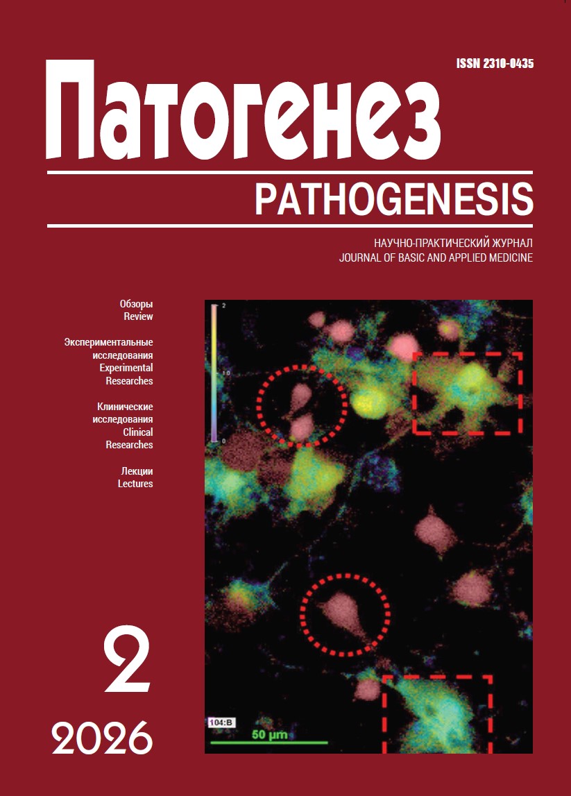
Methods. Neuroglial cultures were obtained from the cerebral cortex of neonatal Wistar rats and used for fluorescence microscopy measurements at 10–13 days in vitro. Changes in the intracellular free Ca2+ concentration ([Ca2+]i) were recorded on a Nikon Ti-2 microscope using the fluorescent indicator Fura-2. To assess the contribution of Ca2+ stored in the ER to the overall Glu-induced increase in [Ca²⁺]i, a calcium-free buffer (Ca2+ replaced with 0.1 mM EGTA) and the endoplasmic Ca2+-ATPase inhibitor thapsigargin (5 μM) were used. AIDA (100 μM) was employed to inhibit mGluRs, and MK-801 + CNQX (10 μM each) to inhibit iGluRs.
Results. When exposed to Glu (100 μM), 90% (7 experiments, 537 cells) of cells responded with a rapid increase in [Ca²⁺]i. In 15% of cells, Glu induced [Ca²⁺]i oscillations, which in Ca2+-free buffer revealed a single [Ca2+]i rise inhibited by thapsigargin, which is typical for astrocytes and indicates the expression of type 1 mGluRs, mGluR5.
Conclusion. Analysis of the morphological features of cells in culture in combination with measurements of Ca2+ signaling using inhibitors of ionotropic and metabotropic glutamate receptors, removal of Ca2+ from the extracellular medium, and inhibition of the ER Ca2+ pump allowed us to evaluate the temporal profile of signals and the ratio of neurons and glial cells in the neuronal culture. A tendency toward a decreased contribution of mGluRs to the Glu-induced increase in [Ca2+]i was found in those cells, likely neurons, which revealed a massive Ca2+ influx from the surrounding buffer. The parameters of the dynamics of [Ca2+]i changes may be useful for testing the neurotoxicity of substances that affect Glu-induced Ca2+ signaling.